Surgery

Select a Category
Dr. Giri performing Laser Surgery
We now offer affordable TPLO Surgery for ACL repair in Dogs.
Here at Rainbow Veterinary Hospital, we have cutting edge surgical equipment, and use the latest surgical techniques. Your pet is in good hands with experienced doctors, and trained technicians.
Laser Surgery
We are pleased to offer laser surgery as an exciting new option for safe, comfortable treatment. Laser technology reduces the trauma to your pet, improves recovery, and often shortens hospital stays. Lasers have been successfully used on humans for over 30 years. This experience is proving to be beneficial for pets and their owners. Ask if your pet's surgical procedure can be done with a laser!
Orthopedic Surgeries
Cranial Cruciate Ligament Injury (CCL Tear)
The most common injury in a dog’s knee joint is a cruciate ligament tear. The cranial cruciate ligament connects the femur to the tibia. The most common cause of CCL (ACL) tears in dogs and cats is degeneration of the ligament and arthritis. When the CCL (ACL) is torn the knee joint is destabilized and pets may suffer lameness and pain.
Diagnosis of CCL (ACL) Tear
Physical examination: Pets may feel pain on palpation of knee joint, lameness, partial pressure on toes while walking. We complete orthopedic examination followed by x-ray under anesthesia.
How is CCL (ACL) Treated?
Since CCL (ACL) tear is the disruption of the knee joint, surgical stabilization is needed.
TPLO Surgery
A dog’s knee joint has excessive tibial slope compared to a human (flat knee joint). TPLO surgery involves removing the excess slope on the tibia by rotating it and stabilizing it with a locking plate so free slide of femur is reduced and the joint is stabilized.

TPLO Surgery
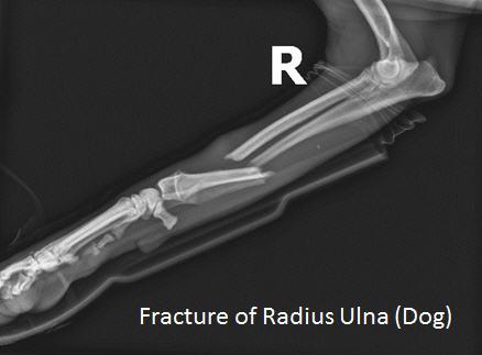
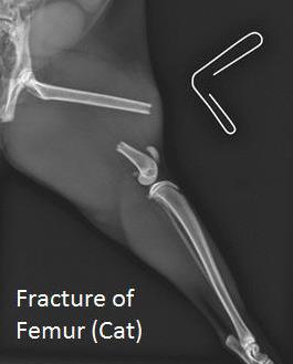
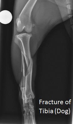
Long Bone Fracture
Pets can get fractures of their long bones due to trauma while playing or unfortunately hit by a car. We offer advanced surgical repair of all fractures of long bones.
Dr. Giri is a member of AOVET and uses AO principles of fracture repair in dogs and cats. Surgical management of the fracture involves the following techniques:
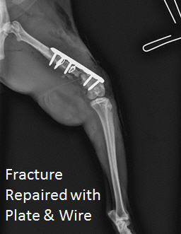
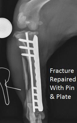
Pinning
Stabilize fracture by various sizes of stainless steel pins and wires across the fracture line.
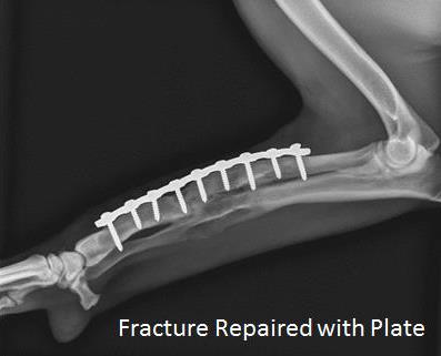
Plating
Fracture is stabilized using stainless steel or titanium plate to the bone using screws on either side of the fracture.
We provide detailed instruction for the physical therapy that will be needed at home. Healing of the bone takes from 6 weeks to 36 weeks depending on the pet’s age and the type of fracture.
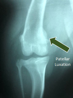
Patellar Luxation
Patella (knee cap) is a small bone in the knee joint that can be dislocated in dogs and cats, which causes lameness, pain, and arthritis.
Patellar luxation is a problem in all breeds and sizes of dogs, but the condition is most common in small breeds of dogs. Commonly affected breeds include the Yorkshire Terrier, Maltese, Toy Poodle, Miniature Poodle, Pomeranian, Pekingese, and Chihuahua.
Clinical Signs
Pet owner typically report a skipping lameness in affected pets. Typically, the pet uses the affected leg normally between skipping episodes. Lameness that worsens in patients with patellar luxation may be associated with a concomitant tear of the cranial cruciate ligament. Cranial cruciate ligament injury occurs in approximately twenty-five percent of patients with patellar luxation.
Causes of Patellar Luxation
1. Shallow trochlear groove
2. Tibial tuberosity displacement
3. Torsional abnormalities of the femur or tibia
Diagnosis
Diagnosis is done based on clinical signs, physical examination, and radiographic findings.
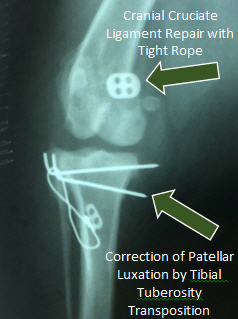
Treatment
Surgical Management
1. Deepening of trochlear groove
2. Tibial tuberosity transposition
3. Corrective osteotomy of femur
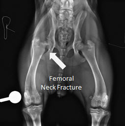
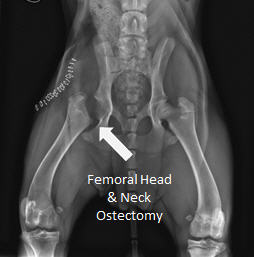
Femoral Head and Neck Ostectomy (FHNO) for Hip Dysplasia and Femoral Neck Fracture
Abnormal development of hip joint in dogs results in hip laxity. This is a genetic condition which causes hip dysplasia. In small, medium, and sometimes large breed dogs, poor fitting ball and socket (hip joint) develop arthritis causing constant pain and lameness.
Sometimes due to trauma, the femoral neck can get a fracture.
Diagnosis of Hip Dysplasia
Physical examination: clinical signs like stiffness, exercise intolerance, and x-ray of hip.
Femoral Head and Neck Ostectomy (FHNO)
To eliminate the pain caused by either the hip joint, laxity, arthritis, or femoral neck fracture, the femoral head and neck is removed. This is a salvage procedure.
Laparoscopy Surgery
Minimally Invasive Laparoscopic Surgery in Natrona Heights, PA. We have performed minimally Invasive Laparoscopic Surgery in Dogs and Cats.
What is Laparoscopy?
Laparoscopy is done by making a small incision and inserting a laparoscope or camera into the abdominal cavity. The abdomen is filled with carbon dioxide (a safe, absorbable gas) to easily see all the organs in the abdomen. The images are magnified and viewed on the monitor in the surgery suite. Structures can be easily examined, and if need be biopsies can be taken. With some procedures no extra incisions may be needed even if additional entries or ports are needed they are only one-quarter to one-half inch incisions.
So how does this affect my pet?
So, what is the big excitement about this advance moving into the veterinary world? Very small incisions decrease postoperative pain, risk of infection, and speedy recovery time. Where a traditional incision to do a liver biopsy would be at least six inches on a 40-50 pound dog, a one-half inch incision can give you excellent visualization of the abdomen and multiple liver biopsies. The camera has magnification so that even the tiniest amount of bleeding can be observed and monitored post biopsy.
Spays involve bluntly breaking down the pet’s suspensory ligament which can be painful. In a laparoscopic assisted spay the ligament is cauterized and then sharply cut. During surgery this is noticeable as there is no/little increase in patient’s heart, blood pressure, respiratory rate. These vital signs often increase during a traditional spay. A recent study in the Journal of the American Veterinary Medical Association showed that laparoscopic assisted spays offered 65% less pain than the traditional open spay.
Another great use for laparoscopy is to do gastropexies, a permanent tacking of the stomach to the body wall. This is a procedure that prevents gastric dilation and volvulus, GDV, a potentially fatal condition that occurs in primarily large deep-chested dogs. Again, the typical incision length would be greater than 6-8 inches, but laparoscopically assisted it can be one to two incisions one-half to two inches in length.
Laser Declawing
Declawing
Cats use their claws to climb and scratch, to defend themselves, and to hunt. Displaying their claws and scratching objects are also considered by many to be a social behavior of our feline friends. Outdoor cats may scratch trees to mark their territory and to remove frayed or worn outer layers from their claws. Unfortunately, this can pose a problem when indoor cats choose their owners’ furniture or curtains as tree substitutes.
What can you do about your cat’s destructive scratching?
A variety of options are available; however, owners often choose declawing as a means to end destructive scratching in the home. Declawing is controversial, as it provides no health benefit to the cat and is done strictly for human benefit. Opponents say it is unnatural and cruel, and can result in psychological damage to the cat. Proponents say that declawing has no more negative effects than does any other surgical procedure, and that by ridding unwanted behavior, it could increase the chances for a cat to enjoy a safe, permanent indoor home. The American Animal Hospital Association is opposed to the declawing of domestic cats unless all other attempts have been made to prevent the cat from using its claws destructively or when clawing presents a significant health risk for people within the household.
Your options
To help caring cat owners decide the best option in their situation, we’ve provided some facts on declawing and on alternative methods that address the problem of destructive scratching by house cats.
Declawing is an irreversible surgical procedure performed by a veterinarian while the cat is under general anesthesia. Hospitalization for one to two days may be required. As the back feet are rarely used for scratching, the front feet are usually the only ones declawed.
A cat’s toe has three bones; the claw grows from the end of the last bone. In declawing, the veterinarian amputates the end section of the last bone, along with the nail. This removes the claw and prevents it from growing back. The toe is then sewn shut with absorbable sutures or closed with surgical skin glue, and each paw is bandaged snugly to control bleeding. Bandages may be removed within one to two days.
Declawed cats require special care immediately after the surgery. Pain medications are often administered for three to five days after surgery. Although difficult to do, owners need to restrict their cat’s activity, especially jumping, for several days. Until healing is complete, the cat should be kept indoors, and shredded newspaper or non-granular litter should be used. Even once the cat has fully recovered, it is wise to restrict him from the outdoors as he really has no adequate means of defense.
Complication rates are very low if the procedure is performed properly. Most cats will walk fairly well within two to three days, although the feet will be tender for about a week or two after surgery. The cat should be seen by a veterinarian if any of these signs occur: swelling, discharge from the toes, loss of appetite or some other change in the cat’s health or behavior. It is normal for a cat to initially limp or favor a paw following surgery. However, make sure to contact the veterinarian if this behavior stops and then resumes again. Additionally, keep aware of bleeding. Although some spotting after surgery may occur and is normal, if bleeding persists, the cat should be rechecked by the doctor.
Laser surgery is another option available for declawing your cat. Surgical lasers have been used for several years at veterinary colleges, but just recently has this technology become an affordable option for veterinary hospitals to offer to clients. A laser declaw surgery requires anesthesia and amputation of the bone and nails (as described above). However, a surgical laser can offer several advantages to a scalpel. As it cuts, the laser automatically seals small blood vessels and nerve endings around the cut, which means less bleeding and less pain. Patients can thus experience a quicker return to their normal activities.
Younger cats tend to recover more quickly and adapt more easily to the loss of their claws.
Most declawed cats will resume normal activities, including performing scratching motions. With rear claws intact, cats can still climb small trees, hunt and even defend themselves when necessary.
Dentistry
Eighty Percent of all dogs and cats 3 years and older have periodontal disease.
This common problem can lead to significant pain, tooth loss, and infection in distant organs such as the heart, liver, and kidneys.
Dental Prophylaxis
Dental prophylaxis is the routine cleaning and polishing that keeps your pet's teeth and gums healthy and prevents offensive breath. Along with regular visits, we offer consultations on home dental care. Having good oral hygiene will allow your pet to enjoy a long and happy life.
Imaging
X-Ray/RadiologyRadiology
X-Ray/RadiologyRadiology is becoming a routine procedure for confirming and diagnosing many diseases in small and large animals. X-Rays are used to contrast densities in order to determine abnormalities. Radiology is very important in veterinary practice due to the frequency of disorders and the increased emphasis placed on obtaining a correct and precise diagnosis.
Ultrasound
Ultrasound is the use of sound waves, much like sonar, to outline organs and tissues in a noninvasive way. We have ultrasound equipment on site to allow us to make accurate and timely diagnoses for your pet.
Video VetScope
The Video Vetscope is a tiny color video camera connected to a probe. It transmits color videos to a monitor. The camera may be used to better diagnose ear infections or blockages, pinpoint dental or throat conditions and/or other problems that might not be seen. It helps the doctor see what's happening and aids in diagnosing diseases.
Laboratory
The health and safety of your pet is our foremost concern. By testing blood chemistries, we can evaluate the status of your pet's major organs and provide an inside look at the blood itself. Testing on a regular basis lets us monitor your pet's health over its lifetime and enables us to detect any potential problems early on.
Telemedicine
"Just one phone call away."
Telemedicine offers the most advanced and complete internal consultations. Medical images taken at our hospital are sent via the internet to specialists around the country. This saves you, the pet owner, time and money over making a separate trip to a far off specialist, where they would likely need to take additional images. Consultants can give our doctors and clients the fastest and most accurate expertise available.
Teleradiology
Teleradiology uses X-Ray scanners and the internet to send X-Ray images to radiologists for interpretation These specialists then send back a diagnosis to our hospital and recommend treatments.
Telesonography
Telesonography captures images directly from the ultrasound device and sends them to a specialist. Where ultrasounds are relatively new to most of the veterinary world, telesonography allows diagnostic images of your pet to be read by Radiologist (Ultrasound) and Cardiologist (Echocardiogram).
Wellness Testing
By age two, most dogs and cats have already reached adulthood. By the time they reach age four, they're considered middle-aged. At the age of seven, many dogs have already started their senior years. Because dogs and cats age five to seven times faster than people, an annual visit to the veterinarian every year is equivalent to people seeing their physician or dentist every five to seven years. People want the best health care for themselves and their pets. And for dogs and cats, that means regular wellness exams every six months.
Is Your Pet A Golden Oldie?
The age at which pets enter their senior or geriatric years depends on body weight. This form was developed to help you determine if your pet is senior or geriatric. On it, you can find the relative age of your pet in human years. For example, a 9-year-old dog weighing 70 pounds is 61 in people years (senior), while a 14-year-old cat is 72.
Best Care for Older Pets
Twice yearly exams help the veterinarian detect, treat, and ideally, prevent problems before they become life threatening. Senior dogs and cats are at higher risk for a number of problems, including diabetes, heart disease, kidney disease and cancer. Many of these conditions are treatable if diagnosed early. That's why it's especially important for older pets to receive a wellness exam twice a year.
Advanced Procedures
We are also capable of performing many advanced procedures to help us diagnose any health problems your pet may be suffering from. Some of the most useful procedures are: X-Ray , Ultrasound , Endoscopy, and Pathology profiles. With all of these resources available to us, we feel confident that we will be able to quickly diagnose and speed your pet on its way back to health.